
 |
|
| C O N T E N T S |
| C-Fern Home |
| Web Manual |
| Introduction C-Fern Nutrient Medium Culture Conditions Gametophyte Culture Gametophyte Observations Sporophyte Culture Spore Mutagenesis Bibliography |
|
Light Stand Growth Pod DNA Extraction C-Fern T-shirt Who's Who Selection and mutants Media formulation error Image gallery |
| Web Journal |
| Educational Resources |
|
National Science Standards Gametophyte development Student research questions Research in the classroom Research with C-Fern Brief description of C-Fern C-Fern manipulation Root tropism? |
| Frequently Asked Questions |
| Workshop and Exhibit Schedule |
| Investigations Mutant & wild type spores Manual & Culture Instructions Supplies - Domes, media |
 |
| Development
of C-Fern is supported by the National Science Foundation (NSF-DUE) |
|
Copyright
© 1997-2000
Thomas R. Warne and Leslie G. Hickok. All rights reserved. |
|
C-Fern Image Gallery
|
|||
|
Click on thumbnail
for larger image |
|||
 |
 |
 |
 |
|
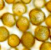
C-Fern
spores.
|
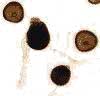
Germinated
C-Fern spores.
|
Germinated
C-Fern spore stained with acetocarmine.
|
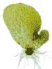
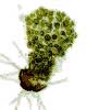
Young
hermaphroditic C-Fern gametophyte.
|
 |
 |
 |
 |
|
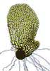
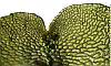
Hermaphroditic
C-Fern gametophyte.
|
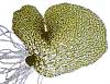
Mature
hermaphroditic C-Fern gametophyte.
|
Mature
hermaphroditic C-Fern gametophyte.
|
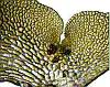
Close-up
of the notch region of a mature hermaphroditic C-Fern gametophyte.
|
 |
 |
 |
 |
|
Close-up
of the notch region of a mature hermaphroditic C-Fern gametophyte.
|
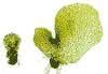
Mature
male and hermaphroditic C-Fern gametophytes.
|
Mature
male and hermaphroditic C-Fern gametophyte.
|
Close-up
of notch region of mature hermaphroditic C-Fern gametophyte stained
with acetocarmine.
|
 |
 |
 |
|
|
Mature
male C-Fern gametophyte.
|
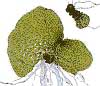
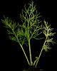
C-Fern
sporophyte with fertile fronds.
|
Close-up
of C-Fern fertile frond showing sporangia.
|
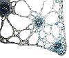
Cross
section of C-Fern petiole showing aerenchyma with large air spaces.
|
 |
 |
 |
 |
|
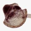
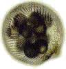
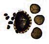
Detached
C-Fern sporangium containing spores.
|
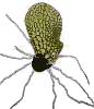
C-Fern spores (on right) are about 120 um in diameter. Woodfern (Dryopteris sp.) spores and sporangium on left and center, respectively. |
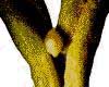
Close-up
of axis of fertile frond showing vegetative bud.
|
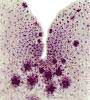
Cross
section of a C-Fern sporophyte bud.
|